ATG5 dans la polarisation des cellules B et la présentation des antigènes
Lien vers l'article originalAuteurs
Florent Arbogast, Johan Arnold, Philippe Hammann, Lauriane Kuhn, Johana Chicher, Diane Murera, Justine Weishaar, Sylviane Muller Icon,Jean-Daniel Fauny & Frédéric Gros
Année de publication
2018
Journal
Autophagy
Résumé de l'article
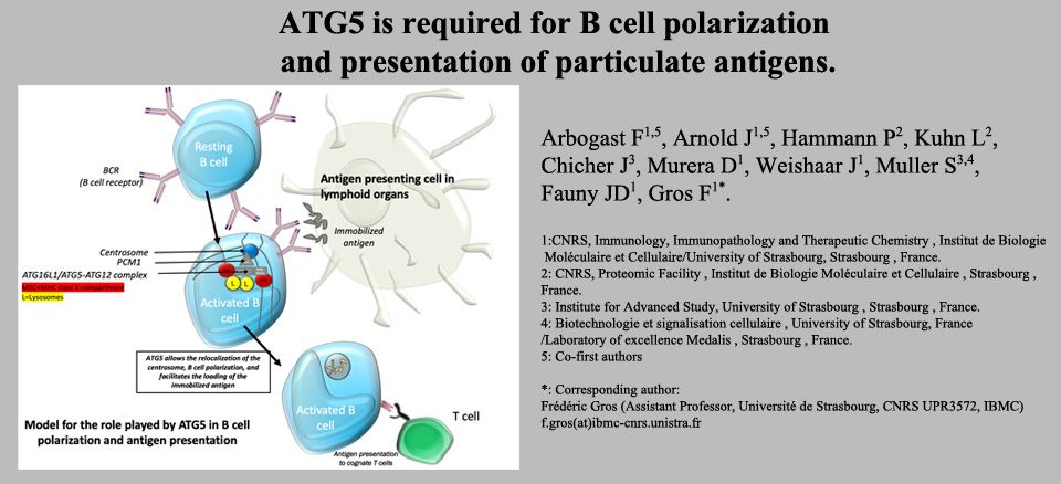
B cells are central actors of the humoral response. Autophagy is implicated in the maintenance of long-lived B cells like antibody-secreting plasma cells and memory B cells. Autophagic activity also allows the presentation of cytosolic antigens by major histocompatibility complex class II (MHC-II) molecules. Antigen presentation is crucial for the crosstalk with T cells, contributing to the terminal differentiation of B cells. Some antigens are presented after capture by the B cell receptor (BCR), the membrane-bound version of the antibody ultimately secreted by plasma cells. The role of autophagy in the presentation of such antigens is suspected but not well understood. Our work shows that ATG5 (autophagy-related 5) allows the colocalization of the BCR with LC3 and ATG16L1 after internalization. Although autophagic machinery is not necessary for the endocytosis of BCR/antigen complexes, ATG5 allows the relocalization of the centrosome at the vicinity of the place where antigen is first internalized and then processed on MHC-II molecules. ATG5 and ATG16L1 are part of a complex comprising PCM1 (pericentriolar material 1), which expression also participates in this phenomenon. This polarization of B cells is major when antigen is immobilized on solid phase, mimicking its availability at the membrane of cells in the follicles of secondary lymphoid organs. In this context, ATG5 optimizes B cell presentation by MHC-II molecules to T cells and thus allows a more effective humoral response. We are currently investigating the interactions between ATG proteins and centrosome components, that promote the reorganization of B cell cytoskeleton, the loading of antigens on MHC-II, and the optimization of the immune response.
Image d'illustration